Imaging
In many cases it is useful to be able to image areas of the body using techniques such as x-rays, ultrasound and endoscopy.
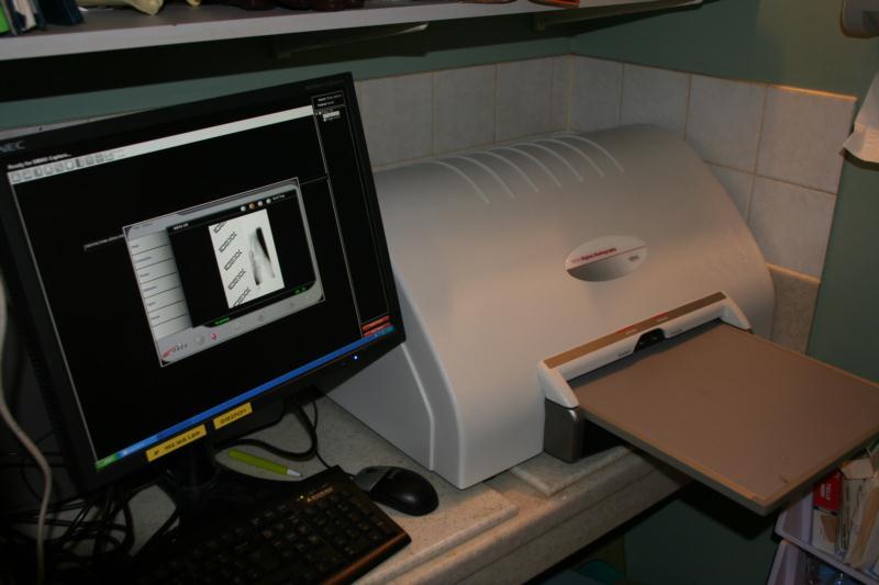
We have a digital x-ray system at both Brampton and Longtown surgeries. Just like digital cameras, digital radiography allows very rapid production of very high resolution images of your pet. This allows quicker diagnosis and the images now are so much more detailed it often allows more subtle changes to be identified. This system also allows us to view x-ray images on high resolution monitors around the Practice (including Branch Surgeries), and also allows us to email the images to consultants .This has dramatically improved the speed at which second opinions can be sought.
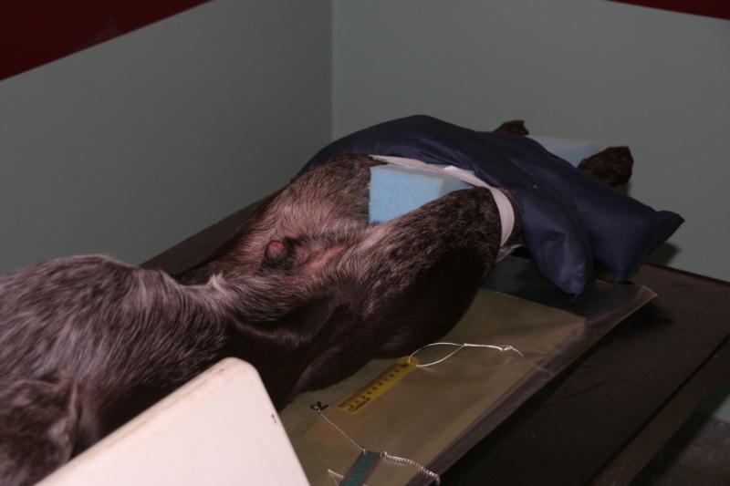
X-rays allow us to image inside the body and are particularly useful for assessing bone and joint problems in orthopaedics. They are also helpful to assess chest and abdominal diseases and to search for hidden cancers. They have limitations in that they can only differentiate between certain tissue types, these being gas, fat, soft tissue, mineralised tissue and metal. The radiograph on the right shows the stifle (knee) joint of a dog. Air can be seen as the black area to the left, bone is seen clearly as almost white (metal appears pure white). The gaps between the muscles (soft tissues) can be seen as there is fat between the muscles (soft tissue appears light grey and fat is a darker grey).
Therefore, if tissues are of the same type and are adjacent, they cannot be seen as separate structures. This can be why foreign bodies that have been swallowed are not always visible on x-ray films. Usually animals need anaesthesia for radiography as they need to be very still.
Ultrasound
Our ultrasound machine is used to see the internal tissues in "real time". This can often be done without sedation. Ultrasound uses high frequency sounds that reflect back to the probe to determine the type of tissue below. It is particularly good at detecting fluid (that appears black on the screen) that accumulates abnormally in some conditions. It can also provide information on the structure of organs such as the heart, liver and kidneys that may show signs of disease. The colour-flow Doppler facility allows us to track the movement of blood within the heart and identify abnormalities. Ultrasound cannot determine the cell type of organs and can therefore only give rise to suspicion of cancers. A biopsy would be required to confirm the actual disease present.
Endoscopy
We have several flexible endoscopes that are all fibre-optic instruments that can be used to look inside such places as the airways, the intestinal tract and the bladder. Small biopsy samples can be taken using the scopes that can provide a diagnosis without the need for major surgery.